Please utilize your web browser's ZOOM feature to
manually optimize the display of this webpage to
your screen resolution

HostSim: a virtual host of infection
|
an on-line supplement for A virtual host model of Mycobacterium tuberculosis infection identifies early immune events as predictive of infection outcomes |
[ Kirschner Lab Time-Lapse Simulations Homepage ]
Supplementary Material
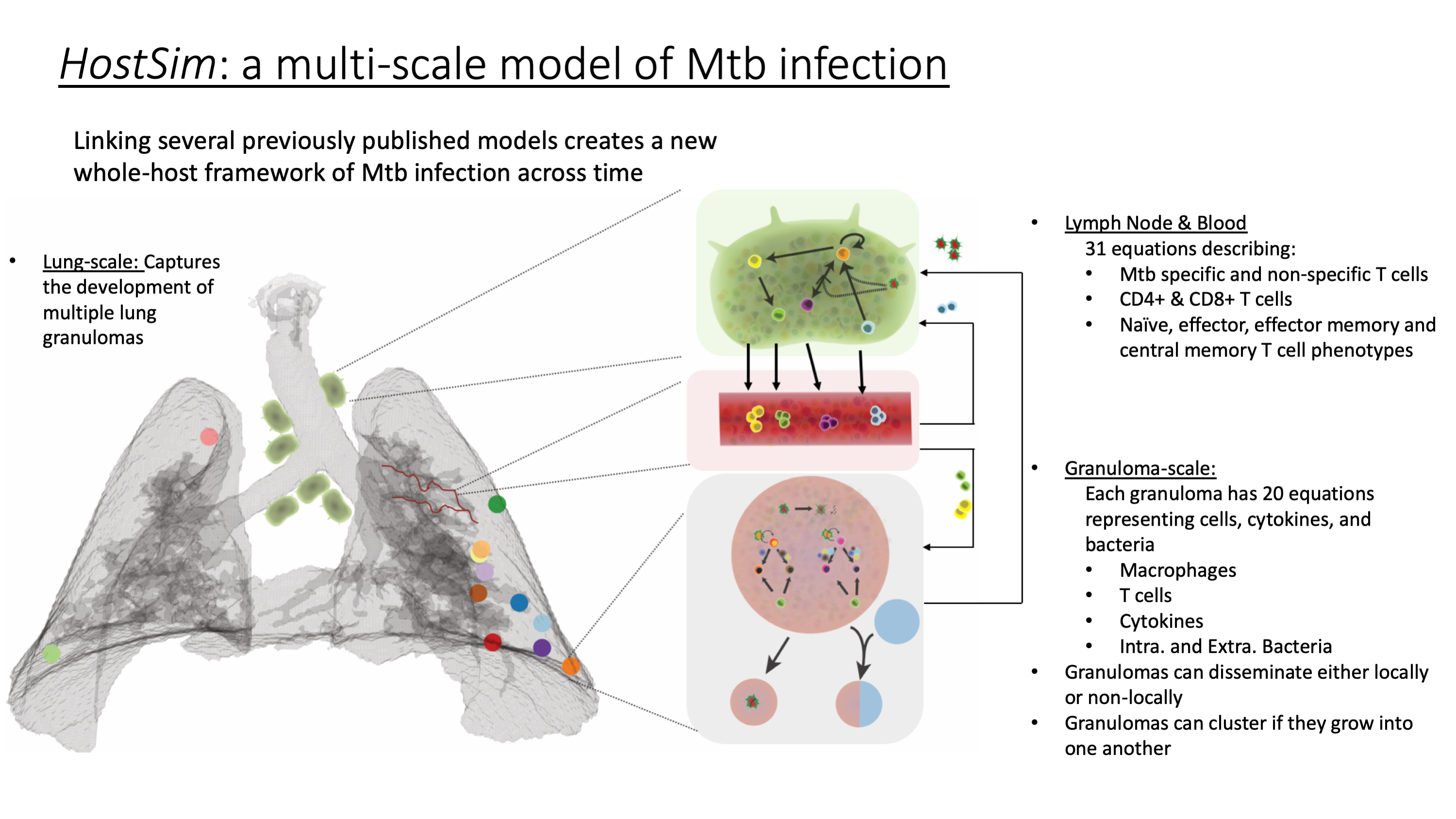

Overview: HostSim, the whole-host computational model of Mycobacterium tuberculosis (Mtb) infection, is a hybrid, multi-scale model that links ordinary differential equations (ODEs) at the cellular and cytokine scale within granulomas to events happening at the whole-host scale, like dissemination of granulomas across lungs and cellular dynamics within the lymph nodes and blood across time.
Within the lungs, each granuloma is modeled as an agent that is placed randomly within the boundary of a 3D lung environment. This 3D lung environment is created from a series of CT scans of a non-human primate (NHP) lung by our collaborators in Dr. JoAnne Flynn’s laboratory at University of Pittsburgh. After granulomas are given a location, each granuloma develops independently across time. Within each of these agents, a system of 20 ODEs track cytokines, immune cells, and bacterial concentrations across time. These ODEs track active, infected and resting macrophages as well as various types of Mtb-specific and non-specific T cells within the granuloma environment. Granulomas can also disseminate, thereby spreading bacteria throughout the lung environment (according to a set of rules). One can think of this model like a metapopulation model, wherein each ‘population’ is a granuloma that interacts indirectly through a lymph and blood compartment model and directly through dissemination events.
We capture blood and lymph node dynamics in a two-compartment model using a series of 31 ODEs that represent T cell priming, proliferation, and differentiation following antigen presentation within the lymph node and blood. Both Mtb-specific and non-specific T cells are tracked across time. This model has been presented before to represent both infection and vaccination dynamics.
HostSim links the lung and lymph node and blood in two main ways. First, a percentage of infected macrophages within the granuloma environment traffic to the lymph node as APCs. This summed influx of cells from multiple granulomas kick-starts the lymph node and blood model. Second, Mtb-specific effector and effector memory T cells are recruited to the granuloma environment. At the end of each timestep (1 day), we calculate the total number of T cells that a granuloma could ideally recruit based on macrophage counts and inflammatory cytokine levels within each granuloma. We sum this total T cell number, N, across each of the granulomas and look within the blood. If the blood has N T cells, then those T cells are allocated to each granuloma within the next timestep. However, if the blood does not have that many T cells (as can be the case early in infection), then the actual number of Mtb-specific Effector and Effector Memory blood T cells is allocated to each granuloma proportionally in a need-based manner. That is, granulomas with more infected macrophages and higher inflammation will proportionally received more T cells from the blood than granulomas with less infected macs/inflammatory cytokines. Additionally, non-specific T cells are recruited from the blood to each of the granulomas. Importantly, T cells in the blood are tracked as a concentration of cells per mm^3, but are converted to counts upon entering the granuloma environment.
HostSim time-lapse video for 200 days showing virtual lungs, granulomas, lymph nodes, and blood cell concentrations for 3 cell types. The blood vessels representing Effector, Effector Memory and Central Memory T cells will change color across time, from black (very few cells in the blood) to bright red (representing the maximum number of cells of that blood type across the simulation). At day 70, Effector T cells have reached their maximum across the simulations and are therefore bright red. Effector Memory T cells are continuing to grow in magnitude, thus the vessel is colored burgundy, and central memory T cells have not yet started to differentiate in large numbers, therefore the Central Memory T cell vessel is colored black, but as the simulation progresses they will become red. Lymph nodes transition from black to green and back to black as antigen presenting cells enter the lymph node in high numbers (green) and then stop entering the lymph node as time progresses. The lung at the left colors the granulomas according to bacterial ID, and you can track granuloma dissemination events by the color of the granulomas. The lung at the left is the same simulation, but colors the granulomas on a range from black:red:yellow, where black means there are no proliferating cells within the granuloma, but yellow indicates that there is a large number of proliferating cells within the granuloma.
HostSim pseudo code: The main solve function within HostSim is outlined in the following:
For i in 1 to the number of simulation days
Pull T cells from the blood, and hold them in matrix, GranulomaRequests
For j in 1 to the number of granulomas
Allocate T cells from GranulomaRequests to each granuloma{j}, by adding the T cell counts to each initial condition
Solve granuloma{j} from i-1 to i timesteps
Save simulation output and timestep output information
Granuloma{j} sends APCs to lymph node
Lymph node receives APCs from granuloma{j}
if (local/non-local dissemination event function == 1) newgranuloma = createNewGranuloma() granuloma{end+1} = newgranuloma; end
end
solve lymph node & blood equations from i-1 to i timesteps
save simulation output and timestep output information
end
|
| [ Kirschner Lab Time-Lapse Simulations Homepage ] |
|
All content and files © 2021 The Regents of the University of Michigan All rights reserved - Kirschner Lab: [ http://malthus.micro.med.umich.edu/lab/ ] |