To preview the movie, click on the graphic image above
Supplement Movie 1
T cell Track in the LN
LN is at a baseline steady state, with CD4+ (red), CD8+ (blue) T cells and
DCs (green) on grid. No Ab-DC is recruited. One of the T cells is tracked
with a yellow line from its entry of the grid from HEV (orange) to its exit
from EL (yellow).
Right click or CTRL-click on AVI icon,
select "Save Link As" or "Save Target As"
to download the archived AVI movie file version of
time-lapse simulations of controlled infection
To extract the AVI movie from the archive, download the archive to your Mac or Windows desktop,
double-click on them, and follow the on-screen instructions.
To extract on Linux, Unix, BSD, etc... use the command in a shell 'unzip (filename.zip)' or a desktop extraction utility
To preview the movie, click on the graphic image above
Supplement Movie 2 The process of T cell priming
This is a illustration of how T cells are primed by DCs in our model. For
clarity, one single Ab-DC (green) is placed at the center of the grid
scanning its Moore neighborhood for cognate T cells. CD4+(red) and CD8+
(blue) T cells recruited from HEVs (orange) moves in the grid following the
rules and bind to the DC if conditions are met. After priming, T cells
unbind and begin to proliferate (pink), and eventually differentiate into
effector cells (purple). Effector cells exit the grid via ELs (yellow).
Right click or CTRL-click on AVI icon,
select "Save Link As" or "Save Target As"
to download the archived AVI movie file version of
time-lapse simulations of TNF Deletion
To extract the AVI movie from the archive, download the archive to your Mac or Windows desktop,
double-click on them, and follow the on-screen instructions.
To extract on Linux, Unix, BSD, etc... use the command in a shell 'unzip (filename.zip)' or a desktop extraction utility
To preview the movie, click on the graphic image above
Supplement Movie 3
Acute infection
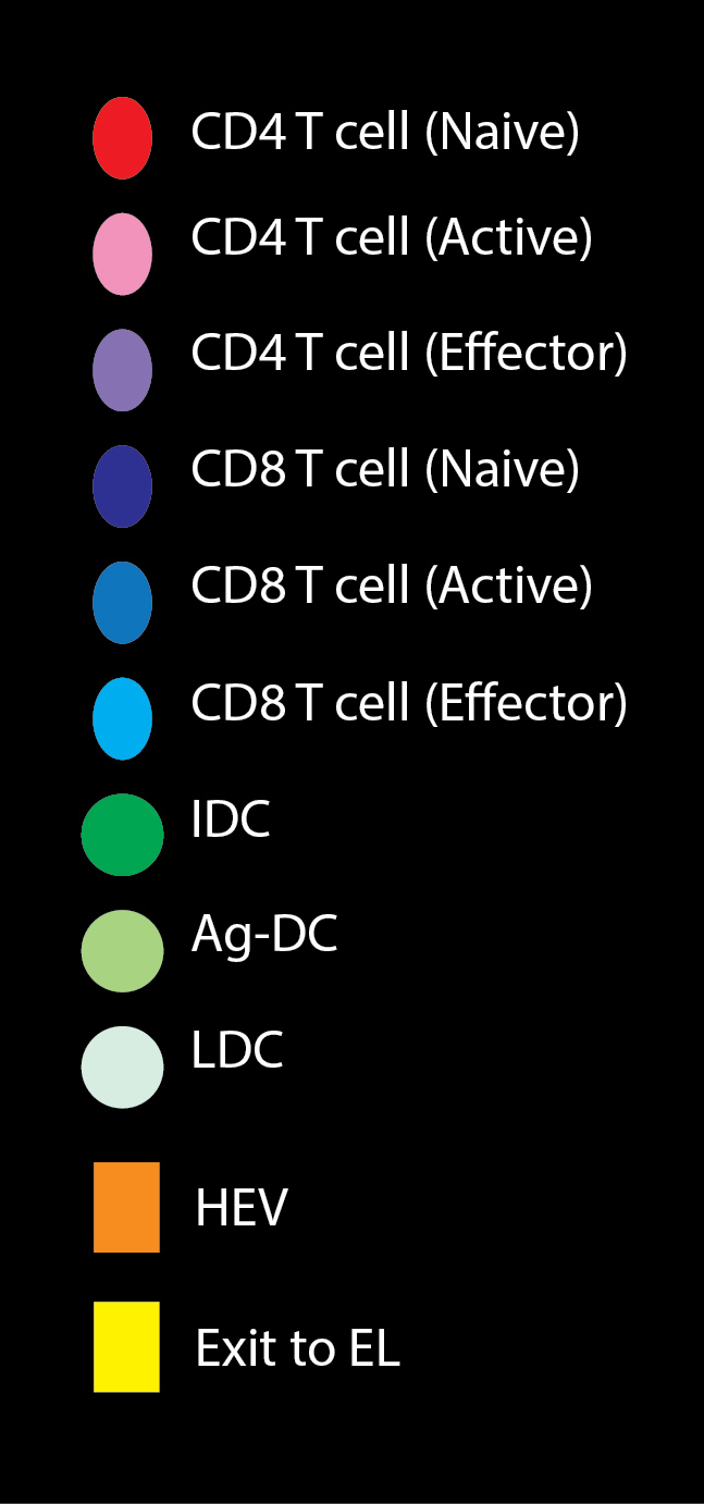
Time-lapse movie of simulated acute infection in the LN. Simulation lasts
for 20 days. Antigen-bearing dendritic cells are recruited from day 5 to day
10. CD4+ T cells: red: naive; pink: proliferating; purple: effector. CD8+ T
cells: blue: naive; sky blue: proliferating; cyan: effector. DC: green:
Immature; light green: antigen-bearing; white: licensed.
Right click or CTRL-click on AVI icon,
select "Save Link As" or "Save Target As"
to download the archived AVI movie file version of
time-lapse simulations of controlled infection
To extract the AVI movie from the archive, download the archive to your Mac or Windows desktop,
double-click on them, and follow the on-screen instructions.
To extract on Linux, Unix, BSD, etc... use the command in a shell 'unzip (filename.zip)' or a desktop extraction utility